Diagnosing And Treating A Cholesteatoma
A cholesteatoma is a destructive, noncancerous growth that expands throughout the middle ear. It begins as a cyst that sheds dead squamous epithelial cells, and these keratinizing, or hardening, cells keep building upon each other accumulating fluids, debris, and sebum. As it grows, it can destroy the small middle ear bones affecting balance, facial muscles, and hearing. Ultimately, it can cause vertigo and especially hearing loss. It is caused by a congenital disability as an abnormal growth inside the ear canal or from repeated inner ear infections. Sometimes, it is caused by the eustachian tubes not properly functioning regulating air flow and pressurization. Learn about how to diagnose and treat a cholesteatoma now.
Hearing Tests

Hearing tests are often the first diagnostic test performed when a cholesteatoma is suspected or confirmed. These tests, performed by an audiologist with an audiometer, are designed to figure out the level of hearing and therefore, the extent of damage from the cholesteatoma. Hearing sensitivity is tested at different frequencies. Patients will wear headphones while short tones are played and respond to which side the tone is heard on, if at all. Another type of test involves repeating words spoken through the headphones at different volumes. These tests can be done in as little as thirty minutes and are painless. Hearing loss occurs with a cholesteatoma when it grows into the delicate ear bones. Though hearing cannot be regenerated once lost, it can be enhanced with hearing aids.
Continue reading to reveal more ways to diagnose and treat a cholesteatoma now.
Computerized Tomography Scan

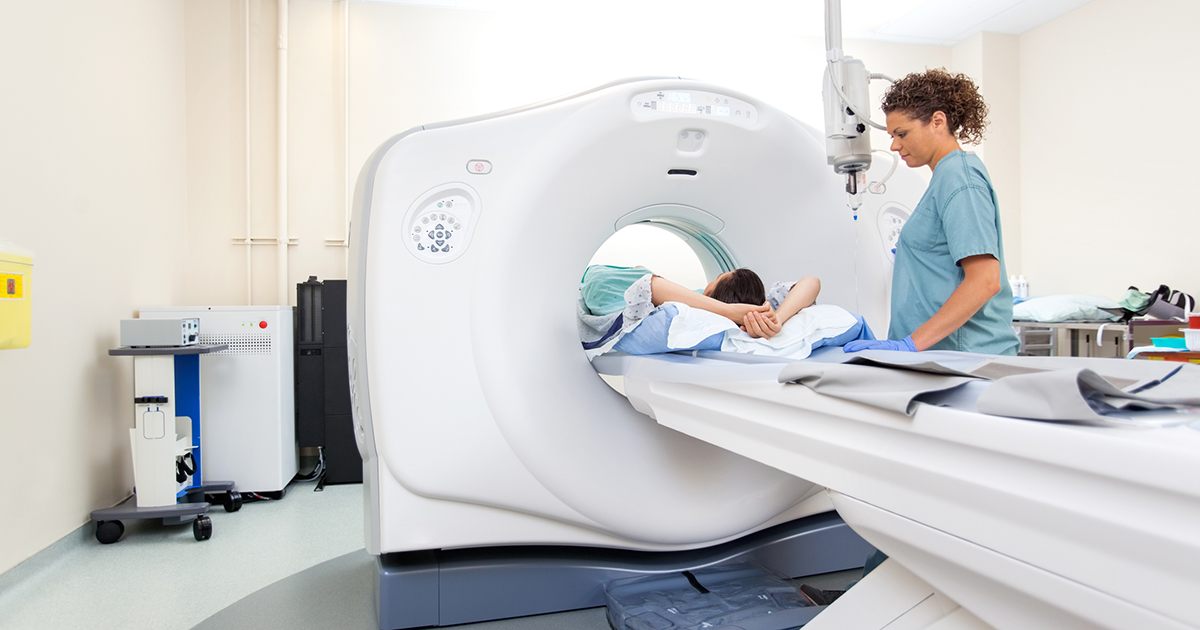
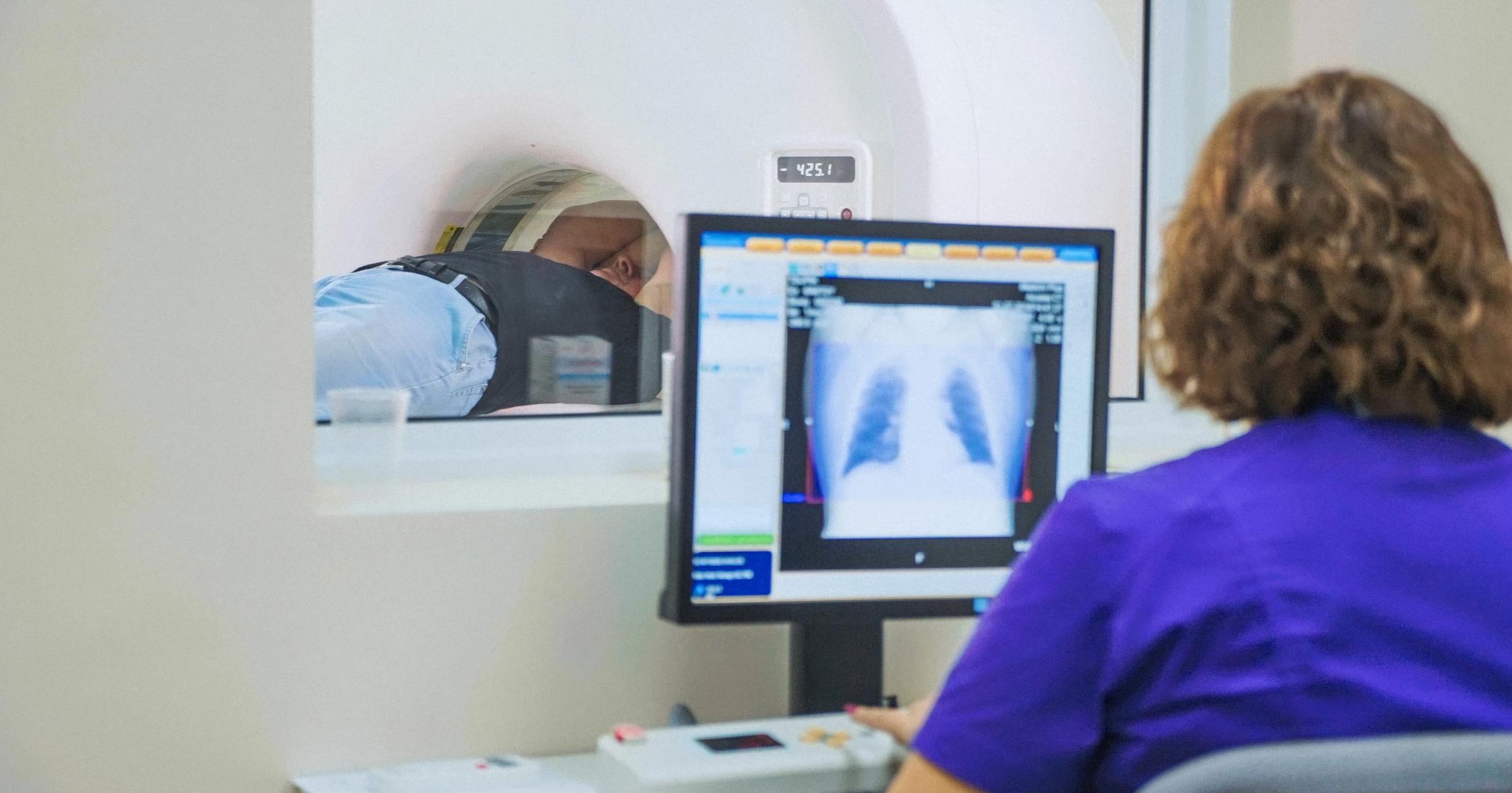
A computerized tomography scan, or CT scan, is a series of X-rays that can determine how far a cholesteatoma has grown into the bones of the ears and mastoid process. Narrow X-ray beams go through the body as the scanner circles around the patient. It takes multiple pictures of the area layered on top of each other to provide more detail than regular X-ray images. This allows for an all-around view of the cholesteatoma, which is especially helpful if the doctor has planned for its removal, as it will show the doctor the amount of bone erosion the cholesteatoma has caused. Though quite detailed, a CT scan cannot often differentiate between scar or inflammatory tissue and cholesteatoma tissue.
For even more detail, magnetic resonance imaging (MRI) may be necessary.
Magnetic Resonance Imaging

If a CT scan determines further imaging is required, magnetic resonance imaging can provide even more detail. Unlike a CT scan, an MRI does not use X-rays instead using radio waves and a magnetic field. This technique is noninvasive and can be especially useful for pre- and post-operation. A person is placed within the MRI scanner, and a 3-D image is produced without the use of harmful radiation. This can show precisely where the excess skin of the cholesteatoma is growing, how deep it has progressed, and help to determine the best way to remove it. Using magnetic resonance imaging post-operation allows doctors to determine if the area is healing properly, has developed an infection, or if scar tissue has formed.
Learn more about treating a cholesteatoma now.
Mastoidectomy

A surgeon performs a mastoidectomy to remove the air cells of the mastoid process, the bone directly behind the ear. This is often the best way to remove a cholesteatoma that continually causes infection. The incision is made in the valley behind the ear, and the cholesteatoma can be removed. Depending on how much it has grown and how much damage has been caused to the inner ear, the bones of the inner ear can be repaired or reconstructed. They can also be replaced with prosthetic bones. The main goal of the surgery is to remove any infection, but often hearing is improved after the cholesteatoma has been fully removed. Complications can include pain and infection, though this is rare. The ability to taste can be altered if the nerve near the ear is affected during surgery.
This is often performed before a tympanoplasty, which is coming up next.
Tympanoplasty

Tympanoplasty is the surgical repair of the eardrum. If the cholesteatoma has grown through the eardrum, it may need to be surgically repaired to improve hearing. This can be performed under anesthesia but also as an outpatient procedure. Different types of repair can be done depending on factors like the patient’s age, as well as the cause, location, and size of the tear. Though most patients will regain their hearing from this surgery, it can cause hearing loss if scar tissue builds up. As with a mastoidectomy, care must be taken to avoid damaging facial muscle nerves, or facial paralysis can occur.