Guide To Pancreatic Cancer Tests
Pancreatic Protocol CT Scan

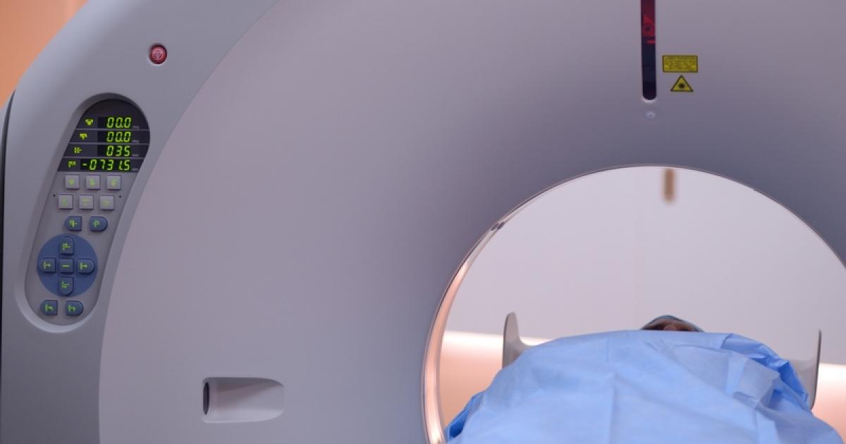
A pancreatic protocol CT scan is a combination of a common diagnostic imaging procedure and a specialized contrast medium administration technique to produce a well-defined representation of a patient's pancreas and surrounding structures. A computed tomography (CT) scan is a machine that takes x-ray images of the interior structures of a patient's body and then utilizes an advanced computer to layer and compound such images into a high-quality three-dimensional representation that can better reveal any odd growths or other abnormalities in the organs. A contrast medium or specialized dye is injected into the patient's circulation to provide better clarity of where the tumor is located relative to the neighboring blood vessels and other organs. When using the pancreatic protocol, the CT scans are taken at specific times during and after the intravenous administration of the contrast medium to make it easier to see abnormalities in the structure, function, and blood flow in the pancreatic structures of pancreatic cancer patients.
Read more about different pancreatic cancer tests now.