How To Effectively Diagnose And Treat Leukodystrophy
MRI And CT Scans

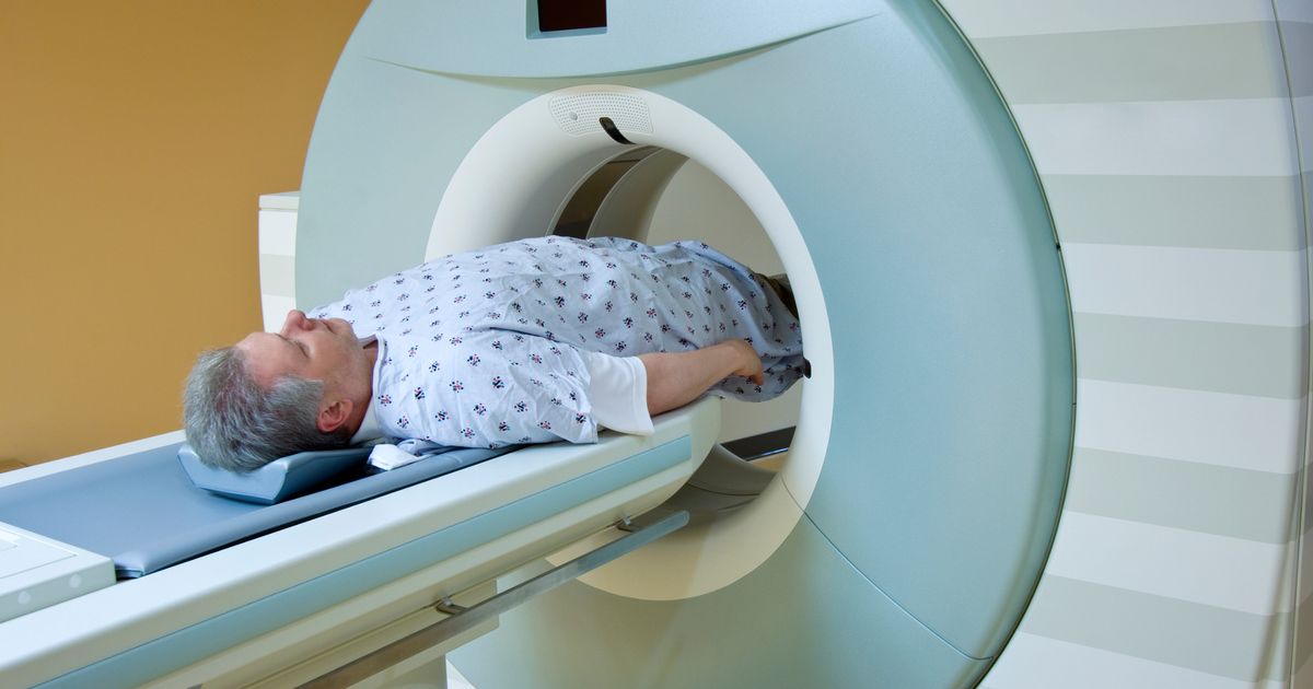
A physician usually uses MRI and CT scans to assist them in making a diagnosis of leukodystrophy. The most characteristic finding on the MRI and CT scans of leukodystrophy patients is symmetrical white matter involvement. The presence of symmetric white matter involvement on an MRI scan indicates a strong possibility the patient has inherited leukodystrophies. Once white matter involvement has been detected on diagnostic imaging scans, the pattern of white matter involvement needs to be identified. There are six different types of white matter patterns on imaging scans, including parieto-occipital, frontal, periventricular pattern, subcortical, brainstem involvement, and cerebellar involvement. Each pattern yields a different leukodystrophy diagnosis. Parieto-occipital patterns are commonly associated with globoid cell leukodystrophy, while frontal patterns are associated with X-ALD and metachromatic leukodystrophy. Periventricular patterns on an MRI are consistent with the presence of metachromatic leukodystrophy, Krabbe disease, leukoencephalopathy with brainstem and spinal cord involvement, and Sjogren-Larsson syndrome. Subcortical patterns indicate L-2-hydroxyglutaric aciduria, and brainstem involvement pattern indicates Alexander's disease.
Get more information on diagnosing leukodystrophy now.