Diagnosing And Treating A Cholesteatoma
Computerized Tomography Scan

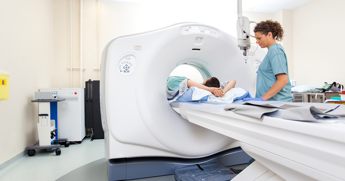
A computerized tomography scan, or CT scan, is a series of X-rays that can determine how far a cholesteatoma has grown into the bones of the ears and mastoid process. Narrow X-ray beams go through the body as the scanner circles around the patient. It takes multiple pictures of the area layered on top of each other to provide more detail than regular X-ray images. This allows for an all-around view of the cholesteatoma, which is especially helpful if the doctor has planned for its removal, as it will show the doctor the amount of bone erosion the cholesteatoma has caused. Though quite detailed, a CT scan cannot often differentiate between scar or inflammatory tissue and cholesteatoma tissue.
For even more detail, magnetic resonance imaging (MRI) may be necessary.