How To Diagnose And Treat A Strawberry Hemangioma
MRI Or CT Scan

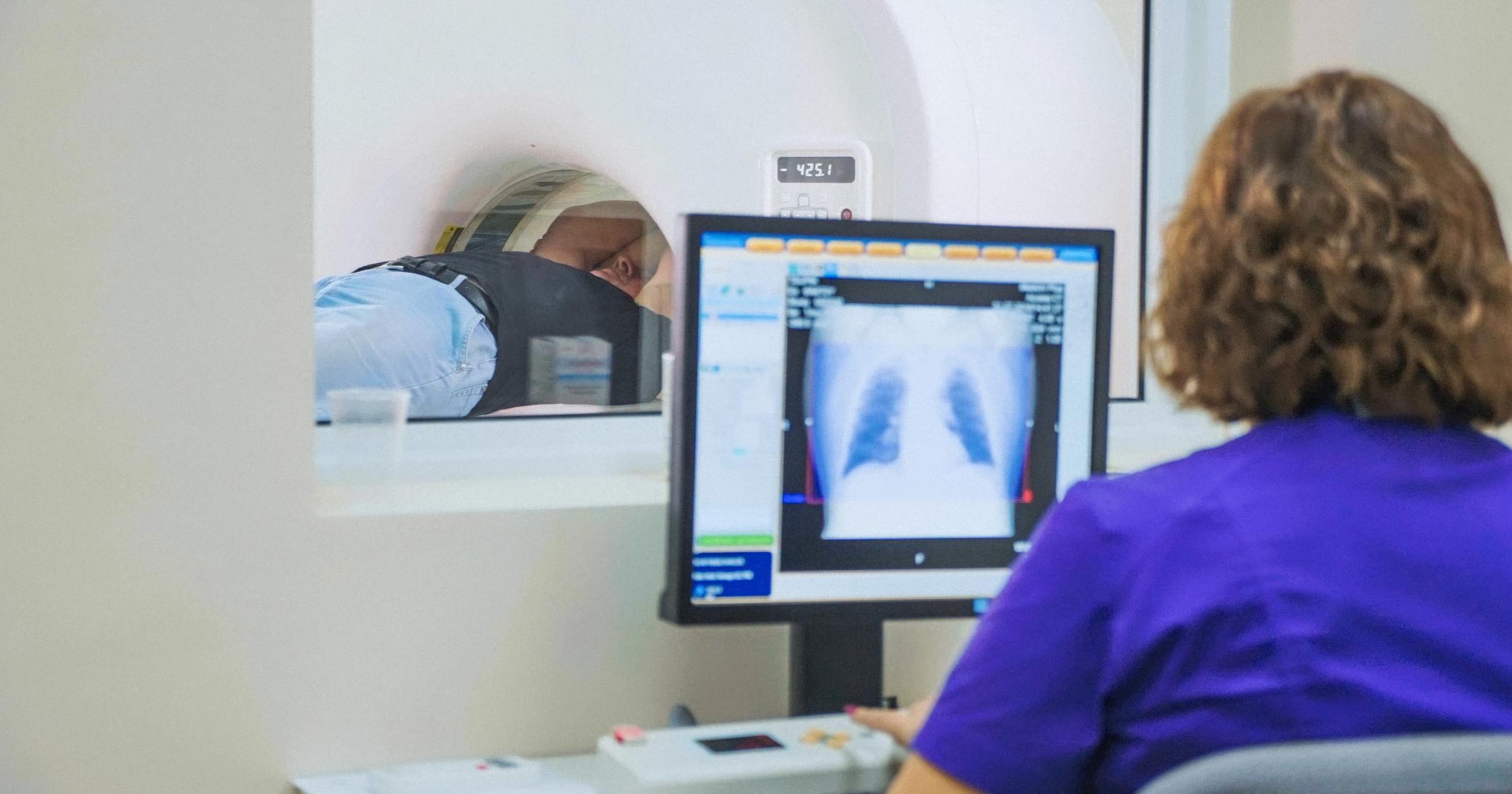
Patients who have large strawberry hemangiomas that may threaten a vital organ typically need to have an MRI or CT scan. These imaging studies are performed by radiologists, and they help doctors get a more detailed view of the depth of the birthmark and find out more information about the blood vessels that may be involved. CT scans use radiation for imaging, and MRI scans use magnetic fields to produce images. Doctors will recommend a particular type of scan after assessing the patient's overall health. Both tests are painless, and the patient simply lies on a table while the imaging device works above them. If a patient is anxious, they may be given a mild sedative in advance of the procedure. Sometimes, the doctor may ask for the scan to be completed with and without contrast. For scans completed with contrast, the patient either drinks a solution or is injected with a substance that enables the area in question to be seen more clearly on the scan. MRI and CT scans can be particularly useful if surgery to remove a hemangioma has been recommended.
Learn about how to treat a strawberry hemangioma next.