How Does An X-Ray Work?
X-rays are some of the most common imaging tools in medicine. They can be used to detect and diagnose a great deal of different bodily issues, from broken bones to blocked blood vessels and cancer. But how do x-rays work? You might have heard they use radiation to create their images. Being nervous about the use of radiation in medicine is natural. But x-ray technology has become safer and safer over the years, with qualified technicians and researchers helping to minimize the risks. X-rays are an essential part of being a medical professional, and they're also an essential element to diagnose many conditions. It's crucial to have a full understanding of what an x-ray is, and how the technology works.
What Is An X-Ray?

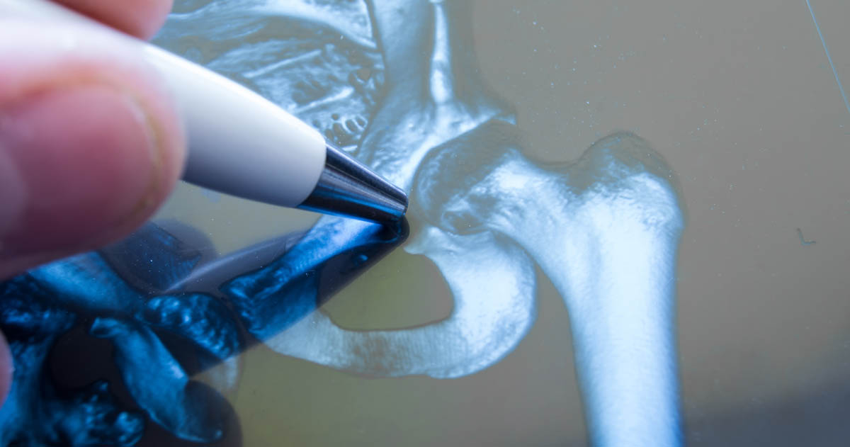
An x-ray is a type of electromagnetic radiation, similar to the light we can see visibly. However, unlike light, x-rays have higher amounts of energy, which allows them to pass through the majority of objects, including your body. A medical x-ray is used to generate pictures of structures and tissues throughout the body. When an x-ray that travels through the body passes through a detector on the patient's other side, an image forms to represent the 'shadows' created by objects found inside the body. There are many different x-ray detectors. One type is photographic film, and others can be used for digital image production. When people capture x-ray images through this process, they are referred to as radiographs.
Continue reading to learn about how x-rays work.